


 will be held in Guangzhou, Shanghai, Chengdu and Beijing")


Researchers from Telethon Kids Institute and The University of Western Australia have developed a new technique to see inside cells with unprecedented detail, revealing a complicated web of interactions that provides new insights into how cells stay healthy.
“For families affected by mitochondrial disease it really opens the way to hope – something they haven’t really had in the 35 years since these diseases were initially discovered,”Professor Aleksandra Filipovska, Lou Landau Chair in Child Health Research at Telethon Kids Institute and UWA
According to lead researcher Professor Aleksandra Filipovska, Lou Landau Chair in Child Health Research at Telethon Kids Institute and UWA, the knowledge gained could pave the way for new treatments for a range of currently incurable diseases – in particular, debilitating mitochondrial diseases which affect up to 1 in 5,000 babies born in Australia.
Much like our body needs organs to function, each of our cells has inner ‘organs’ called organelles. Within each cell, these organelles collaborate, with each performing specific functions: the mitochondria produce energy, the rough endoplasmic reticulum makes and folds proteins that are exported from the cell, the Golgi apparatus processes proteins and fats, and the peroxisome deals with the destruction of fats no longer needed by the cell.
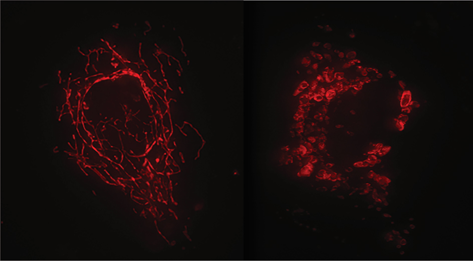 Image: Normal mitochondria networks vs disrupted mitochondria networks.
Professor Filipovska said it was already well known that the structures and functions of organelles in cells depended on each other for cell health, however, until now these relationships had not been systematically explored.
To understand more about how their interactions influence cellular health, she and her team used advanced cellular biology techniques to visualise organelle structure and function in three dimensions.
In a multi-year study detailed today in the prestigious journal Nature Cell Biology, they used a powerful imaging technique – focused ion beam scanning electron microscopy – to watch what happens within cells deliberately engineered to harbour mutations that damage organelles.
“We showed that if one of the organelle team members isn’t doing their job, it can cause trouble for the whole cell – and that has implications for how diseases may be understood and treated,” Professor Filipovska said.
In particular, organelles rely on specific types of fats (ether-glycerophospholipids) to function properly. The study found that when certain genes related to these fats were turned off in cells, it caused problems in various organelles.
“These gene changes led to a decrease in specific fats in the cells, affecting the structure and function of mitochondria – the cell’s powerhouses,” Professor Filipovska said.
“Additionally, disrupted fats impacted how different organelles communicated and behaved. Certain cells, when lacking these fats, showed issues in their Golgi, a structure involved in processing fats. This led to changes that affected overall cell health.”
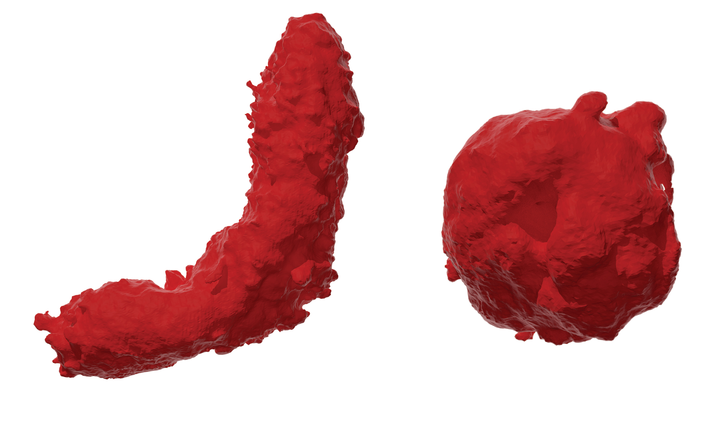 Image: Normal mitochondria vs disrupted mitochondria.
The study explored potential solutions and found that by providing the cells with specific fat-building blocks, they could partially fix the issues.
“Our work has identified that specific lipids can rescue the function of the powerhouses in the cell and improve their communication with the rest of the cell,” Professor Filipovska said.
“This finding has important implications for treatment of diseases caused by diminished energy supplies.”
Professor Filipovska said the study’s findings had the potential to make a huge difference to clinicians’ ability to diagnose disease and could pave the way for therapies for mitochondrial diseases and other diseases caused by cellular mutations, which have lacked effective treatments until now.
“For families affected by mitochondrial disease it really opens the way to hope – something they haven’t really had in the 35 years since these diseases were initially discovered,” she said.
This work was supported by the Channel 7 Telethon Trust and the McCusker Foundation and was a collaboration with a team of researchers including Professor Oliver Rackham from the Harry Perkins Institute of Medical Research and the Curtin Health Innovation Research Institute at Curtin University.
The work is now set to be expanded into a full research program. Using a newly announced Investigator Grant – awarded to Professor Filipovska by the National Health and Medical Research Council last week – the team will build on the study’s findings to address the prolonged and complex diagnostic process for mitochondrial diseases and develop new treatments.
Title image shows detail of 3D visualisation of cell components: mitochondria in red, Golgi in green, ER in blue and peroxisomes in yellow (vesicles are in brown and endosomes in grey) Credit: Dr Ben Padman, Filipovska group member at UWA/TKI