


 will be held in Guangzhou, Shanghai, Chengdu and Beijing")


Engineers from the University of Waterloo are harnessing artificial intelligence to help doctors better see and control a non-invasive cancer treatment and, in the process, save lives.
Their imaging system will allow for the safer and more effective use of high-intensity, focused ultrasound to destroy a wide range of cancerous, often deadly, tumours.
“We are addressing a huge challenge for focused ultrasound treatment,” explained project leader Moslem Sadeghi Goughari, a research associate in the university’s Department of Mechanical and Mechatronics Engineering. “Our imaging system can tell doctors exactly how much of a cancerous tissue is destroyed. And it’s the first AI-powered ultrasound technique developed for focused ultrasound treatment.”
Focused ultrasound treatment uses high-frequency sound waves to deliver a strong beam that heats — and kills — cancer cells. In use since the 1970s, it has been used to treat prostate, breast, liver and other cancers without surgery that requires incisions.
But while focused ultrasound treatment can be done as day surgery and allows for a quicker, more pain-free patient recovery, it has serious drawbacks that limit its widespread use. It is difficult to control with precision which means the treatment can inadvertently damage healthy tissue near a tumour or leave part of a tumour untreated, which allows a cancer to spread.
For Sadeghi Goughari and his three engineering colleagues, accurate monitoring of this therapeutic treatment as it is happening is the key to solving these problems. With no effective enough solution available, they set about developing their own system.

A schematic diagram of Safeghi Goughari’s experimental setup. Photo credit: Moslem Sadeghi Goughari
For hardware, they adopted a focused ultrasound transducer (a device that converts energy from one form to another) to deliver ultrasound energy to a targeted area. They also employed an ultrasound imaging probe to capture images at the area where the ultrasound energy is directed. Given that the area being ablated is tiny — typically in the range of millimetres to centimetres — aligning the ultrasound probe and transducer was critical for proper monitoring. The researchers used a robotic arm to ensure alignment, enabling them to obtain exact images even as the transducer moved during treatment.
For software, they developed an artificial intelligence (AI) framework integrated with the imaging procedure. During treatment, the AI compares the ultrasound images taken before and after the application at an extremely rapid rate. The framework can handle 45 frames of ultrasound images every second, fast enough to be combined with ultrasound imaging systems for real-time monitoring of focused ultrasound treatment. It can detect what happens in the treatment area in less than 22 milliseconds.
Due to this speed, the system can detect how much of a tumour has been destroyed with a 93 per cent accuracy rate. Sadeghi Goughari said this means a doctor could detect “the ablated area margin with accuracy in the range of micrometres.” This could help surgeons better control focused ultrasound treatment, both in terms of destroying a tumor and avoiding damage to healthy tissue.
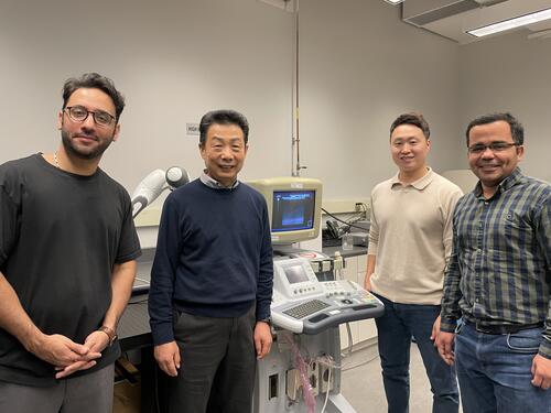
(From L-R) The research team from the Department of Mechanical and Mechatronics Engineering: Moslem Sadeghi Goughari, Research Associate, Dr. HJ Kwon, Associate Professor, Jeong-woo Han, Postdoctoral Fellow and Hossien Rajabzadeh, PhD student.
The team will build on the promising research results by expanding their method and tracking the growth of a treated area in real-time during the treatment.
Medical specialists have given the technology positive feedback with interest from Dr. Jung Suk Sim, Medical Director at the Withsim Clinic in South Korea, to adopt the system for clinical use.
More information about this work can be found in the research paper, “Artificial-intelligence-assisted ultrasound-guided focused ultrasound therapy: a feasibility study,” which appears in the International Journal of Hypothermia.